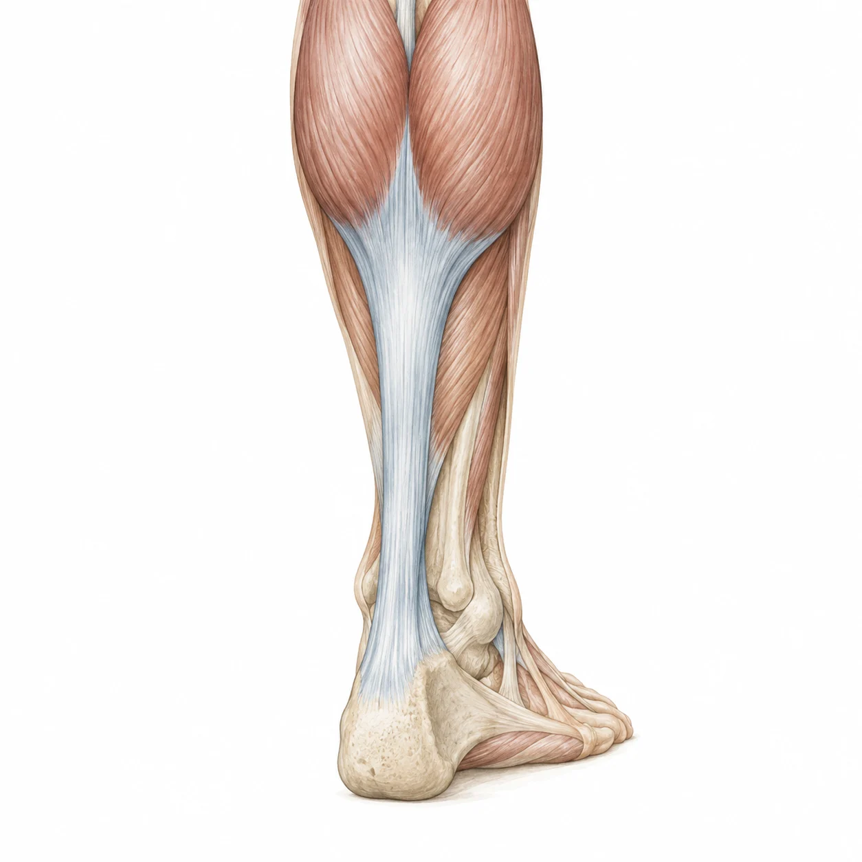
Thickness is one observation between multiple
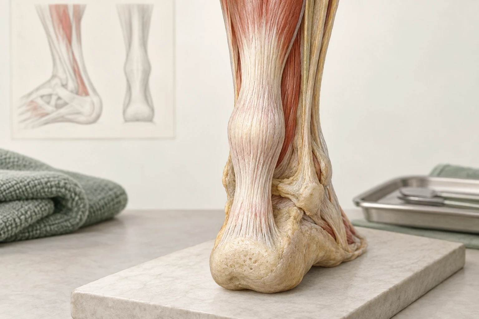
You can notice for yourself that part of the Achilles tendon feels wider, or read the word thickening in an ultrasound report. This can cause unrest: has the tendon been damaged, inflamed or strengthened? The size alone does not answer those questions. The time of onset, the pain spot and what you can use it requires a meaningful interpretation.
The guideline on diagnostic image finding calls thickness in addition to structure and vascularization. These are descriptive characteristics in imaging. They need to be connected to the clinical story. A report with many technical words is therefore not automatically an assessment of the seriousness of your limitations.
Which an ultrasound can and can't clarify
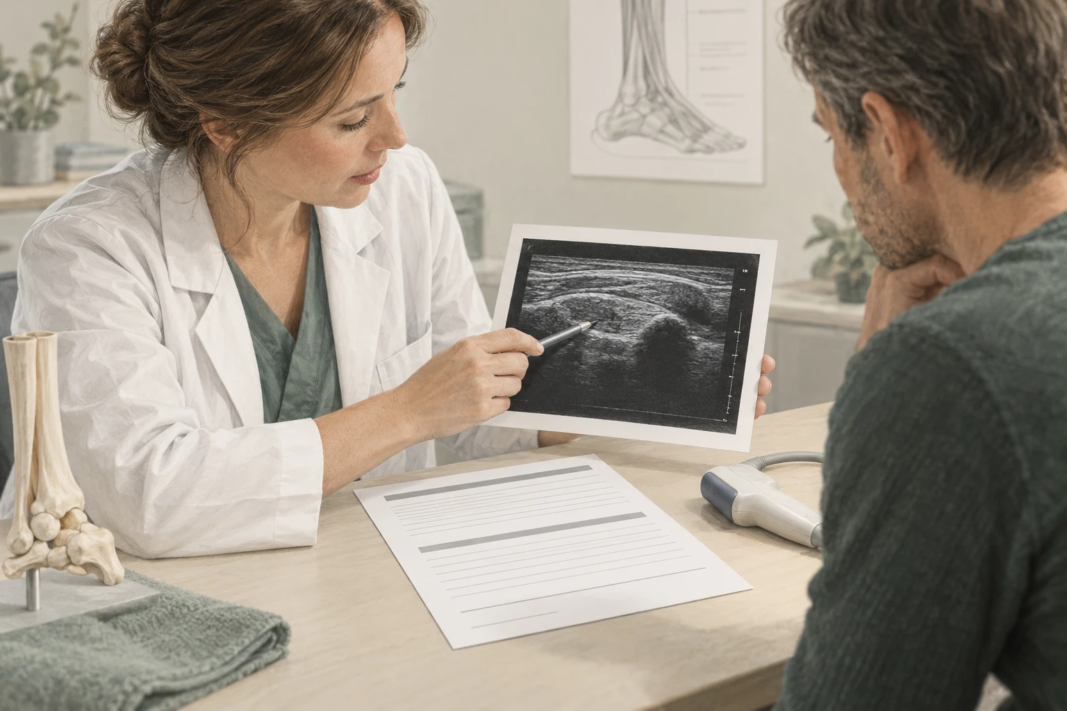
An ultrasound can help to investigate a specific question, for example when the story or findings do not clearly match the probable diagnosis. A picture is made of certain tissues at that time. The rash should be explained in conjunction with study and symptoms, not as a loose report for the tendon.
Ask which part of the result is important to the plan. An abnormal structure without appropriate clinical demand does not need to lead to intensified treatment. Small differences in measurement between studies also require interpretation. The position, measuring site and technique can influence the comparison. Without that context, one millimeter more or less could be mistakenly seen as evidence of deterioration or recovery.
The scan does not predict your recovery date
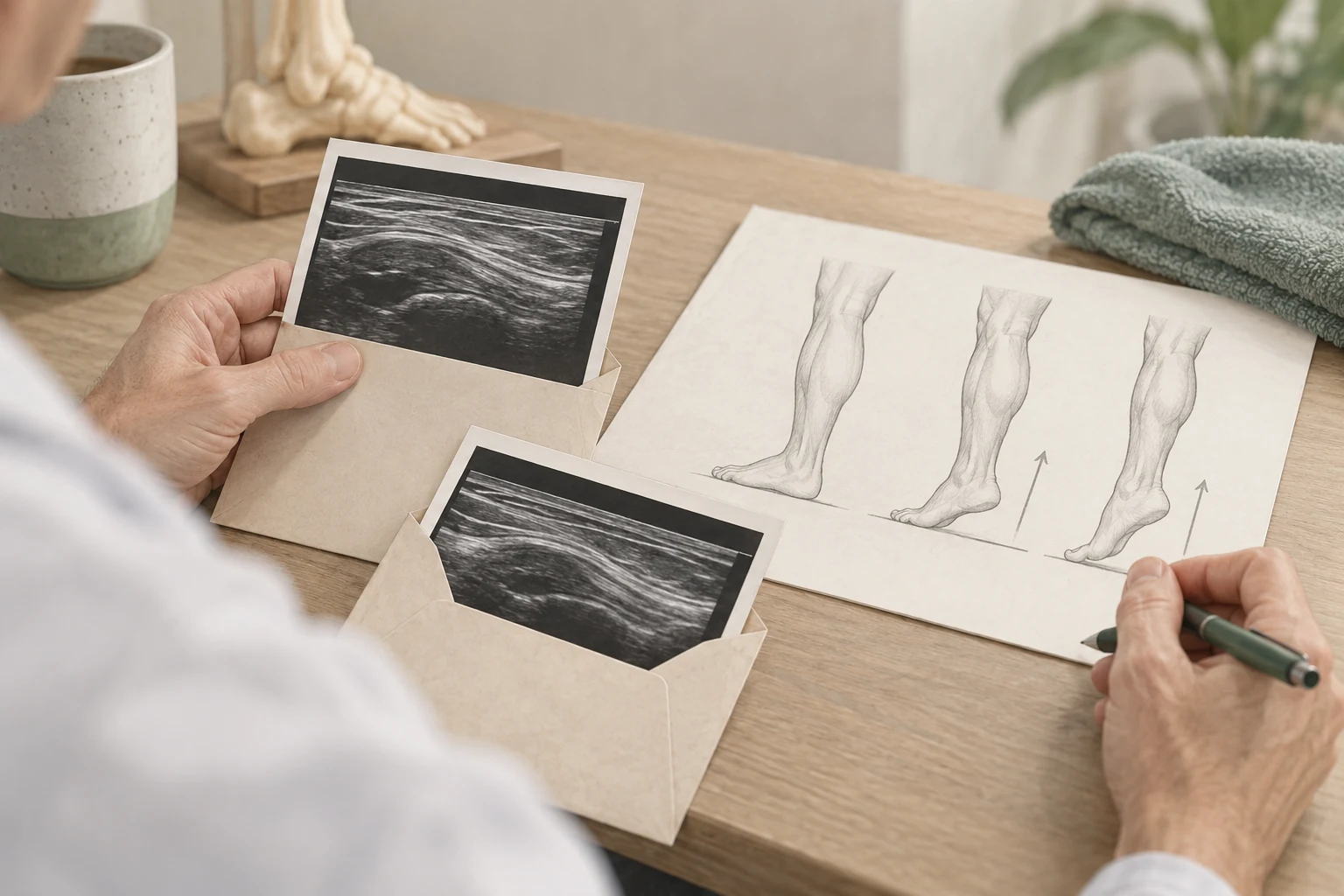
The guideline on prognostic imaging factors advises not to perform imaging to predict the course of symptoms. The available findings do not provide a reliable basis for this. A thicker tendon does not necessarily mean a longer personal recovery time.
That also has implications for goals. Waiting for the tendon to look completely normal can be less useful than following or walking, dropping off and improving the chosen sports load. One example: the visible thickening changes little, while someone travels a daily route more easily. This functional improvement deserves attention, even without spectacular change on the scan.
Feeling constantly can cloud the demand
When you're worried, it's understandable that you often pinch or compare with the other side. As a result, you do not automatically collect reliable information. The sensitivity can change and press repeatedly the spot can make the spot stand out. Moreover, both legs are not always exactly the same, even without symptoms.
Choose which change you want to follow in advance. For example, whether the known thickening suddenly changes, whether new heat or redness develops, or the function decreases. Let the meaning of this be assessed when necessary. For daily recovery a fixed activity often gives more information than the tendon test several times a day between your fingers.
A result interview in five questions
These questions help to move from an image description to an understandable plan:
- Does the place of thickening fit my recognizable symptoms?
- What finding changes our choice for treatment or investigation?
- What words only describe what the tendon looks like?
- How do we monitor functioning if the thickening remains visible?
- What change would be the reason for a new assessment?
Also ask whether repeated imaging is really necessary. A next scan may be useful for a new question, but routine measurement to find reassurance does not always give the desired answer. A clear evaluation target can provide more guidance: what do we want to be able to do, what is the current level and when will the approach be discussed again?
New swelling is something other than a known thickening
A longer-standing, assessed thickening of a tendon is not the same as a sudden swelling after an incident. In case of a handsome, acute loss of marketing power or rapidly changing symptoms, immediate medical assessment is necessary. Also striking heat, redness, sensational changes or unexplained marked increase should be reported.
In the case of stable, longer-standing symptoms, the treatment can continue to focus on appropriate load and functional progress. The healthcare provider should be able to explain why the thickening is relevant or not. Thus, the appearance of the tendon becomes one part of the information, without determining how much confidence you may have in each step or which treatment would be necessary automatically.
Frequently asked questions
Does a thickened tendon have to get thinner before I can work out?
Not automatically. Sport resumption is assessed on symptoms, function, response and the requirements of the activity. Thickness alone doesn't predict that reliably. Let us discuss what functional steps are relevant for your purpose and what role a possible scan has in it.
Is a thickening the same as a tear?
No. It's different descriptions and thickness alone doesn't show a crack. In the event of sudden injury, good-looking or loss of marketing power, it must be assessed immediately whether a rupture is possible. Do not use appearance or feel as a home test to rule that out.
Why is there blood flow in my ultrasound report?
Vascularisation is one of the characteristics that can be described in research. The finding shall be made in the case of symptoms and other information. Ask what significance it has in your situation; only its presence does not give a reliable personal prognosis.
Does every check need a new ultrasound?
No. An ultrasound should answer a relevant question. Often progress can be followed with conversation and functional assessment. A new or unexpected change may give rise to additional research, but routine scanning is not the same as careful evaluation.
Sources and evidence
- guideline Achilles tendinopathy — Diagnostic imaging findings (2020) ↗
Thickness, structure and vascularisation as described image characteristics.
- guideline Achilles tendinopathy — Prognostic Image Factors (2020) ↗
Do not use imaging to predict the expected course of symptoms.
A personal follow-up step
From insight
to an appropriate plan.
At a paid intake, we'll discuss your symptom and do targeted research. ultrasound is included at no extra cost when it makes sense. Any treatments are agreed separately.
View the initial consultation